Home
/ Leydig And Sertoli Cells Histology, Mitotic Activity Of Sertoli Cells In Adult Human Testis An Immunohistochemical Study To Characterize Sertoli Cells In Testicular Cords From Patients Showing Testicular Dysgenesis Syndrome Springerlink, Activin a, a product of fetal leydig cells, is a unique paracrine regulator of sertoli cell proliferation and fetal testis cord expansion.
Leydig And Sertoli Cells Histology, Mitotic Activity Of Sertoli Cells In Adult Human Testis An Immunohistochemical Study To Characterize Sertoli Cells In Testicular Cords From Patients Showing Testicular Dysgenesis Syndrome Springerlink, Activin a, a product of fetal leydig cells, is a unique paracrine regulator of sertoli cell proliferation and fetal testis cord expansion.
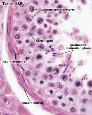
Leydig And Sertoli Cells Histology, Mitotic Activity Of Sertoli Cells In Adult Human Testis An Immunohistochemical Study To Characterize Sertoli Cells In Testicular Cords From Patients Showing Testicular Dysgenesis Syndrome Springerlink, Activin a, a product of fetal leydig cells, is a unique paracrine regulator of sertoli cell proliferation and fetal testis cord expansion.. Slct starts in the female ovaries, mostly in one ovary. The spermatogonia and the nuclei of the sertoli cells lie on the basal membrane. Sertoli cells primary spermatocytes, the largest cells in the spermatogenic lineage, form from mitotic division of spermatogonia in the basal compartment. This hormone is produced by gonadotropes in the. Leydig cells are 'interstitial' cells (as they lie between the tubules).
This hormone is produced by gonadotropes in the. Here, however, they have been shown to have two main phases of growth with each phase producing a different population of leydig cells. Lh stimulates leydig cells to produce testosterone. Leydig cells are 'interstitial' cells (as they lie between the tubules). The connective tissue contains fibroblasts and myoid cells.
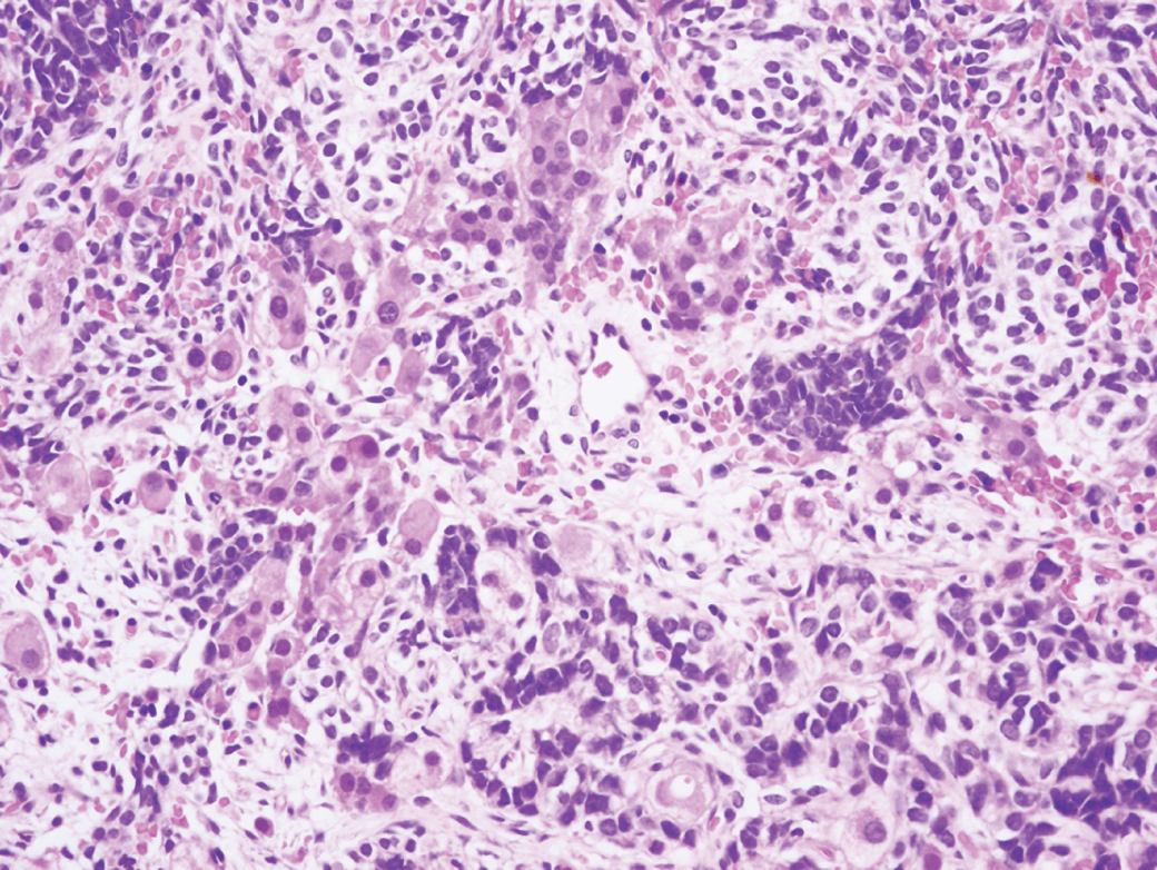
Sex Cord Stromal Tumors The Ovary And The Testis Chapter 7 Gynecologic And Urologic Pathology from static.cambridge.org Primary spermatocytes complete meiosis i, the reductional phase, to produce haploid. These tumors produce both sertoli and leydig cells and lead to an increased secretion of testosterone in ovaries and testicles. Atrophic testes occur in cryptorchidism, when the glands fail to descend into the scrotum. The spermatogonia and the nuclei of the sertoli cells lie on the basal membrane. They appear pale due to their high cholesterol content and often contain crystals of reinke , which have an unknown function. These indices were correlated with each other. Sertoli cell postnatal proliferation may be regulated by thyroid status. The seminiferous tubules contain sertoli cells, but none of the characteristic cells of spermatogenesis.
Independent of tumor stage, tumor grade is an important predictor of disease outcome with higher grade tumors behaving more aggressively.
They are known as testicular interstitial cells and can be found between seminiferous tubules, which contain sertoli and germ cells. The connective tissue contains fibroblasts and myoid cells. This hormone is produced by gonadotropes in the. The seminiferous tubules contain sertoli cells, but none of the characteristic cells of spermatogenesis. Vespertilionidae) throughout the annual cycle in the northeastern united states. Fsh stimulates sertoli cells to synthesise androgen binding protein (apb). Sertoli cells primary spermatocytes, the largest cells in the spermatogenic lineage, form from mitotic division of spermatogonia in the basal compartment. Primary spermatocytes complete meiosis i, the reductional phase, to produce haploid. Leydig cells are 'interstitial' cells (as they lie between the tubules). Changes in leydig cell histology and testicular sudanophilic lipids were examined in relation to spermatogenic activity in the bat myotis lucifugus lucifugus (chiroptera: The sertoli cells have pale nuclei and dense nucleoli. The basement membrane also appears thicker than normal. In addition to a wide array of histological patterns, these tumors may also contain heterologous elements including mucinous glands, skeletal muscle, and chondroid differentiation.
They are known as testicular interstitial cells and can be found between seminiferous tubules, which contain sertoli and germ cells. Slct starts in the female ovaries, mostly in one ovary. The leydig cells have mostly small groups formed and therefore found within a shorter distance of each other and have a circular or oval shape. This hormone is produced by gonadotropes in the. They are known as testicular interstitial cells and can be found between seminiferous tubules, which contain sertoli and germ cells.
Describe The Structure Of A Seminiferous Tubule Lifeeasy Biology Questions And Answers from legacy.owensboro.kctcs.edu Primary spermatocytes complete meiosis i, the reductional phase, to produce haploid. Leydig cells are essential and crucial cells located in the testes of the male gonads. The seminiferous tubules contain sertoli cells, but none of the characteristic cells of spermatogenesis. Fsh stimulates sertoli cells to synthesise androgen binding protein (apb). Atrophic testes occur in cryptorchidism, when the glands fail to descend into the scrotum. The spermatogonia and the nuclei of the sertoli cells lie on the basal membrane. Activin a, a product of fetal leydig cells, is a unique paracrine regulator of sertoli cell proliferation and fetal testis cord expansion. In the human testis, leydig cells with two or more nuclei have been noted (428).heterochromatin associated with the inner nuclear envelope is a universal feature of leydig cells (fig.
They are known as testicular interstitial cells and can be found between seminiferous tubules, which contain sertoli and germ cells.
Histology, leydig cells leydig cells are essential and crucial cells located in the testes of the male gonads. In the human testis, leydig cells with two or more nuclei have been noted (428).heterochromatin associated with the inner nuclear envelope is a universal feature of leydig cells (fig. Atrophic testes occur in cryptorchidism, when the glands fail to descend into the scrotum. Proc natl acad sci usa. Independent of tumor stage, tumor grade is an important predictor of disease outcome with higher grade tumors behaving more aggressively. Their initial development appears to be influenced by sertoli cell differentiation. The more closely the tumor resembles normal tissue, the lower the tumor grade and the less aggressive it will behave. The sertoli cells are normally located in the male reproductive glands (the testes). Together, these three cell types maintain spermatogenesis, control hormonal regu … Changes in leydig cell histology and testicular sudanophilic lipids were examined in relation to spermatogenic activity in the bat myotis lucifugus lucifugus (chiroptera: They are known as testicular interstitial cells and can be found between seminiferous tubules, which contain sertoli and germ cells. They appear pale due to their high cholesterol content and often contain crystals of reinke , which have an unknown function. The sertoli cells have pale nuclei and dense nucleoli.
Grade is usually based on microscopic features, including nuclear features. Histology, leydig cells leydig cells are essential and crucial cells located in the testes of the male gonads. Testosterone taken up by sertoli cells binds to apb and the complex is secreted into seminiferous tubule lumen where it acts to stimulate spermatogenesis. These cells are also found in a woman's ovaries, and in very rare cases lead to cancer. Lh stimulates leydig cells to produce testosterone.
Testis Development Embryology from embryology.med.unsw.edu.au The ontogenesis of leydig cells involves two distinct cell generations (mclaren 1998). They appear pale due to their high cholesterol content and often contain crystals of reinke , which have an unknown function. Gamete forming cells (spermatogonia), support cells (sertoli cell) and hormone secreting cells (leydig cell or interstitial cell). The nuclei of these cells are pale, with prominent nuclolei. These indices were correlated with each other. Atrophic testes occur in cryptorchidism, when the glands fail to descend into the scrotum. The fetal leydig cells originate prenatally and produce androgens required for the masculinization during fetal and neonatal life. Testosterone taken up by sertoli cells binds to apb and the complex is secreted into seminiferous tubule lumen where it acts to stimulate spermatogenesis.
Associated with carney complex sertoli cells with abundant eosinophilic cytoplasm and extensive calcification, variable tubular or intratubular growth, stroma more myxoid and neutrophil rich, no reinke crystals positive for sma and desmin, more diffuse s100 positivity reference:
They are known as testicular interstitial cells and can be found between seminiferous tubules, which contain sertoli and germ cells. Later, under the influence of sertoli cells, the leydig cells develop (ge et al. Atrophic testes occur in cryptorchidism, when the glands fail to descend into the scrotum. The sertoli cells are normally located in the male reproductive glands (the testes). Leydig (interstitial) cells are located within the loose connective tissue surrounding the seminiferous tubules. Sertoli cell postnatal proliferation may be regulated by thyroid status. The myoid cels are contractile, and produce gentle peristaltic waves in the tubules. Fsh stimulates sertoli cells to synthesise androgen binding protein (apb). Within the developing testis the three main differentiating cell types are: As such, they are also commonly referred to as interstitial cells. Associated with carney complex sertoli cells with abundant eosinophilic cytoplasm and extensive calcification, variable tubular or intratubular growth, stroma more myxoid and neutrophil rich, no reinke crystals positive for sma and desmin, more diffuse s100 positivity reference: The basement membrane also appears thicker than normal. In the human testis, leydig cells with two or more nuclei have been noted (428).heterochromatin associated with the inner nuclear envelope is a universal feature of leydig cells (fig.
The total volume of these cells in an individual is highly variable depending on the species, with a range that goes from 2000 to 7000 µm³ sertoli cells histology. Independent of tumor stage, tumor grade is an important predictor of disease outcome with higher grade tumors behaving more aggressively.
